What constitutes a thorough echocardiogram?
Our Protocol
When assessing a patient, there is a minimum set of images that should be obtained in order to allow a thorough evaluation. An ideal study contains both still images and loops, with basic measurements included through post-processing. This includes 2D, m-mode, Spectral Doppler, and Color Doppler modalities. A simultaneous ECG is considered ideal, although is not mandatory for image interpretation.

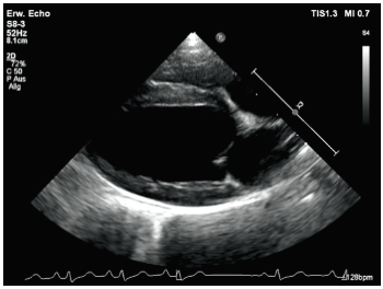
The right parasternal long axis
-
4 chamber – RA, RV, LA, LV
-
5 chamber including the aortic valve/LVOT
-
Color flow on the mitral valve
-
Color flow on the aortic valve
-
Color flow on the tricuspid valve and CW Doppler if TR present
-
In congenital cases, color on the interatrial septum and the perimembranous IVS


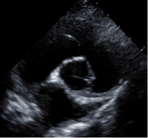
Right parasternal short axis
-
LV, 2D and m-mode
-
Base view for LA/Ao
-
Tricuspid valve
-
Color of the tricuspid valve
-
Base view with PA/RVOT outflow
-
Color of PV/RVOT
-
Doppler – PW and CW of PV/RVOT

Left parasternal
-
Left apical 4 and 5 chamber
-
Color left apical 4 and 5 chamber
-
Doppler of LVOT, especially cats
-
RV/tricuspid valve
-
Color RV tricuspid valve

Left cranial
- RV/TV (ideally if there is suspicion for a tumor)
- Color TV and Doppler if TR present

